
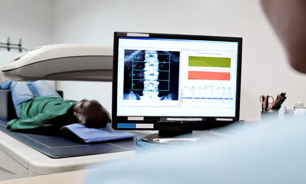
Sixty years ago, the only real way to confirm bone loss was to watch someone break a bone. That’s not a dark joke. That’s the clinical reality before modern densitometry existed. A plain X-ray could only flag bone loss after 30 to 40 percent of bone mass had already vanished, which meant the window for early action was basically nonexistent.
The story of how the field went from that bleak starting point to today’s radiation-free scanning is one of the more underappreciated arcs in modern medicine’s history. It touches on nuclear physics, university labs, a regulatory shift in the 1980s, and, most recently, a technology from Italy that processes ultrasound signals in ways that weren’t computationally possible a generation ago.
The Crude Beginning: Plain Radiographs and Their Obvious Problem
Through most of the mid-twentieth century, physicians relied on conventional radiographs to get any sense of skeletal density. The method was simple, widely available, and fundamentally flawed for this specific purpose. Bone simply doesn’t show meaningful change on a plain film until the loss is severe. By the time a radiologist flagged something alarming, a patient had usually already suffered a fracture or was on the edge of one.
The clinical frustration with this limitation pushed researchers toward a better approach. Single-photon absorptiometry emerged in the 1960s, developed largely through research at the University of Wisconsin, and marked the first time clinicians could actually measure bone mineral at a specific site rather than estimate it visually. Research by the University of Wisconsin in the late 1960s and early 1970s led to the commercial launch of a stand-alone bone densitometer, according to the American Association for Medical Instrumentation. The patient had to sit with a limb immobilized for roughly ten minutes, but it was a genuine step forward in measurable precision.
The problem was that single-photon systems only worked reliably on peripheral sites like the wrist. They couldn’t assess the hip or spine, which are the sites where fracture risk matters most.
The DXA Era: A Leap Forward That Defined a Generation
The real turning point came in the late 1980s. Dual-energy X-ray absorptiometry, known as DXA, landed commercially and almost immediately reset the clinical standard. In 1987, Hologic developed the first bone densitometry device using its proprietary DXA technology, establishing a performance benchmark that would dominate the field for the next three decades.
DXA works by directing two X-ray beams at different energy levels at the patient’s skeleton. By comparing how each beam attenuates through bone versus soft tissue, the system calculates bone mineral density with meaningful precision. The patient reclined on a table rather than holding a limb still. The hip and lumbar spine could both be assessed. Results came back as a T-score, a standardized number comparing the patient’s density against a young adult reference population.
This was a genuine leap. But DXA carries ionizing radiation, requires a clinical referral in most settings, and involves bulky, fixed equipment. For routine screening of an aging population at scale, those aren’t trivial limitations. According to a 2021 NCHS Data Brief from the CDC, the age-adjusted prevalence of osteoporosis among U.S. adults aged 50 and over was 12.6%, with rates among women reaching 19.6%. That’s an enormous population that needs regular monitoring, not a single diagnostic snapshot.
The infrastructure gap between how many people need bone density tracking and how many can realistically access repeated DXA scanning has been a persistent problem in preventive care. DXA solved the accuracy problem. It didn’t solve the access problem.
The Diagnosis Gap Nobody Talks About
Here’s the uncomfortable reality that the DXA era didn’t fix: most people with bone loss still don’t know they have it. A 2025 study published in PubMed using NHANES data found that 69.12% of individuals with osteoporosis went undiagnosed, with the highest undiagnosed rates among men and adults in the 50 to 59 age group, where the condition is easily missed before obvious symptoms emerge.
This isn’t a failure of the technology. DXA is accurate. It’s a failure of reach. When scanning requires a physician referral, a covered diagnosis code, a fixed radiology suite, and a follow-up appointment to get results, a predictable portion of the at-risk population never gets in the door. The undiagnosed majority isn’t skipping screenings because they don’t care. They’re skipping because the friction is high and the urgency feels abstract until a fracture makes it concrete.
The substantial and increasing prevalence among certain groups, along with the lack of diagnostic capture, highlights existing gaps in public health efforts and care delivery infrastructure. (PubMed Central, 2025, analyzing NHANES data from 2005 to 2018)
That gap is exactly what the next wave of scanning technology set out to close.
REMS: The Third Era of Bone Density Scanning
If single-photon absorptiometry was the first era and DXA defined the second, then Radiofrequency Echographic Multi-Spectrometry is best understood as a third-era technology built to solve the access problem without sacrificing accuracy.
REMS uses ultrasound rather than ionizing radiation. A handheld probe acquires unfiltered echographic signals at the hip and lumbar spine. Those raw signals are then analyzed in the frequency domain and compared against reference models to calculate bone mineral density. The key distinction from older ultrasound attempts is that REMS doesn’t just measure signal transmission speed. It processes the full spectral content of the reflected signal, which carries far more structural information.
The clinical validation behind REMS is substantial. A European multicenter study published in the journal Bone found that both REMS sensitivity and specificity exceeded 90% when compared against DXA as the reference standard, according to Contemporary OB/GYN’s 2020 reporting on the research. The Pearson correlation between DXA-measured and REMS-measured bone mineral density was above 0.93 across both assessment sites, a correlation level that clinicians take seriously.
The practical implication is that a provider offering an Echolight REMS bone density scan in MN can deliver accuracy that rivals the hospital-based gold standard without radiation exposure, without a referral requirement, and without the logistical overhead of a fixed radiology suite. For someone who wants to know their bone status and doesn’t want to wait six weeks for a referral appointment to materialize, that matters.
A Practical Lens: The Three Eras Side by Side
| Era | Technology | Decade Introduced | Radiation? | Axial Sites? | Access Model |
|---|---|---|---|---|---|
| First Era | Single-Photon Absorptiometry | 1960s | Yes (low) | No (peripheral only) | Clinical referral required |
| Second Era | DXA | 1980s | Yes (low) | Yes | Clinical referral required |
| Third Era | REMS | 2010s | No | Yes | Direct access, no referral |
What This Means If You’re Due for a Scan
Understanding this history changes how you think about your options. DXA is still excellent and still the standard of care in clinical diagnosis. But it isn’t the only accurate choice anymore, and for routine monitoring or initial screening, the constraints of the DXA model can push important decisions further into the future than they should be.
If you’re over 50, have a family history of fractures, or are entering a period of life where skeletal changes accelerate, here’s a practical way to think about it:
- If your goal is a one-time diagnostic scan ordered by your physician following a fracture, DXA in a clinical setting is appropriate.
- If your goal is regular monitoring of bone health progress over time without radiation exposure and without navigating a referral, REMS is worth knowing about.
- If you’ve been told you have osteopenia and want to track your bone density every six to twelve months, the radiation-free aspect of REMS becomes practically relevant at that frequency.
The field has moved far from the days when a broken hip was the first data point. Six decades of engineering and clinical research produced options that didn’t exist when your parents were your age. Using them is the straightforward part. Knowing they exist is the harder first step, and that’s exactly why the history of this technology deserves more attention than it usually gets.
What would change about how often people got screened if bone density checks were as accessible as a blood pressure reading? That’s the question the third era of scanning is quietly trying to answer.

Comments are closed.